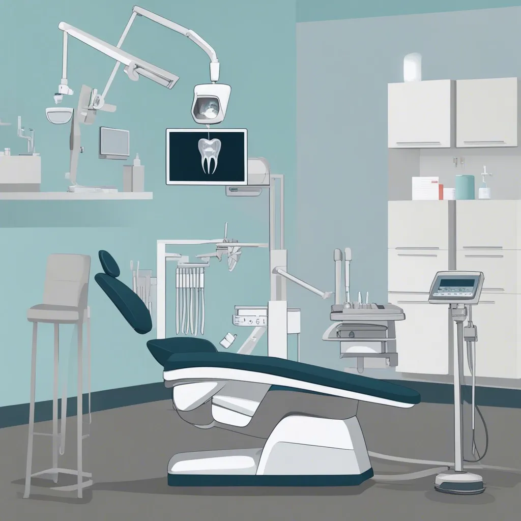
Dental X-Rays Imaging – Understanding the Importance
Dental X-rays imaging plays a crucial role in the field of dentistry, providing valuable insights into oral health and guiding treatment decisions. Among the various techniques used, dental X-rays are a fundamental tool used by dentists worldwide. Dental X-rays, also known as radiographs, are valuable diagnostic tools that allow dentists to visualize the teeth and surrounding structures that are not visible to the naked eye.
In Kusadasi Turkey, dental imaging services, including dental X-rays, are offered to patients as part of comprehensive dental care. These images provide a detailed view of the teeth, bones, and soft tissues, helping dentists detect and diagnose dental problems such as tooth decay, gum disease, and abnormalities. The field of dental radiography, or dental radiology, focuses on utilizing these imaging techniques to ensure accurate diagnoses and effective treatment plans. By capturing high-quality images, dental imaging enables dentists to provide tailored treatments for their patients, improving oral health outcomes.
Exploring Different Types of Dental Radiography
Dental radiography plays a vital role in the field of dentistry, providing valuable information for diagnosis and treatment planning. With the advancements in technology, various types of dental scans are now available to dentists, enabling them to obtain precise and detailed images of the oral structures. Dental X-ray machines are commonly used in dental practices to capture dental X-ray images, which are essential for identifying dental problems such as tooth decay, gum disease, and bone loss.
One of the most common types of dental radiography is digital dental X-rays. This technology has revolutionized the way dental images are captured and stored. Digital dental X-rays offer several advantages over traditional film X-rays, including reduced radiation exposure, instant image availability, and the ability to enhance and manipulate the images for better analysis. In addition to digital dental X-rays, panoramic X-rays are also frequently used in dental clinics.
These dental X-rays provide a comprehensive view of the entire mouth, including the teeth, jaws, sinuses, and surrounding structures. Furthermore, periapical radiographs are another important form of dental radiography that focuses on capturing images of individual teeth and their supporting structures. These detailed images help dentists identify issues such as abscesses, root fractures, and changes in bone density.

The Advancements in Digital Dental X-Rays
Digital dental x-rays have revolutionized the field of dentistry by offering numerous advancements in imaging technology. With traditional film-based radiography, dentists had to wait for the film to be developed and processed before the images could be viewed. However, the introduction of digital radiography has eliminated the need for film, making the process faster and more efficient.
One of the significant benefits of digital dental x-rays is the ability to obtain precise and detailed images in a shorter span of time. Dental x-ray equipment equipped with digital sensors allows for the generation of high-quality intraoral and extraoral radiographs. These digital images can be viewed immediately on the computer screen, enhancing the dentist’s ability to diagnose and treat patients accurately. Moreover, digital radiography allows for the integration of various imaging modalities, such as cone beam computed tomography (CBCT), which provides three-dimensional views of the teeth, bones, and surrounding structures, aiding in the planning of complex dental procedures.
An Overview of Panoramic X-Rays
Panoramic x-rays, also known as orthopantomograms, are a widely used type of dental imaging technique. These x-rays provide a comprehensive view of the entire mouth, including the teeth, jaws, and surrounding structures. Panoramic x-rays are particularly useful in assessing the dental and skeletal relationships, as well as detecting any abnormalities or pathologies in the oral cavity.
Unlike other dental x-ray techniques that require the use of intraoral dental x-ray sensors or films, panoramic x-rays utilize a specialized machine that rotates around the patient’s head. This allows for a single exposure to capture a wide range of dental and facial anatomy. The resulting image can then be used for dental x-ray interpretation and analysis.
When performing a panoramic x-ray, dental professionals must adhere to specific dental x-ray techniques to ensure accurate results. The patient’s head position, along with the machine settings, must be carefully calibrated to capture the desired areas of interest. Additionally, proper dental x-ray exposure settings should be selected to minimize radiation exposure while maintaining image quality.
Overall, panoramic x-rays offer a valuable tool in dental diagnosis and treatment planning. The ability to visualize the entire dental arch in a single image allows dental professionals to assess various dental conditions and determine the most appropriate course of action. However, it is important to note that panoramic x-rays may not be suitable for detecting certain dental issues, such as cavities in between teeth. In such cases, additional dental x-ray techniques, such as bitewing x-rays, may be necessary for a comprehensive evaluation.
The Role of Periapical Radiographs in Dental Diagnosis
Periapical radiographs play a crucial role in dental diagnosis, aiding dentists in assessing the health of teeth from root to crown. These radiographs provide valuable information about the supporting structures of the teeth, including the bone and surrounding tissue. By capturing detailed images of individual teeth and their surrounding areas, periapical radiographs enable dentists to identify a wide range of dental issues that may not be visible during a routine dental examination.
The dental X-ray diagnosis facilitated by periapical radiographs allows dentists to detect conditions such as dental caries, abscesses, cysts, or bone loss. This type of radiography helps dentists evaluate the alignment of teeth and the presence of any impacted or extra teeth. Through a detailed analysis of dental X-ray findings, dentists are able to formulate an accurate dental X-ray report, which plays a vital role in determining the appropriate treatment plan for patients. Moreover, with advancements in dental X-rays technology, periapical radiographs can now be captured with minimal radiation exposure, ensuring dental X-ray safety while providing valuable diagnostic information.
Exploring the Benefits of Cone Beam Computed Tomography (CBCT)
Cone Beam Computed Tomography (CBCT) has revolutionized dental imaging and has become an increasingly popular tool in modern dentistry. CBCT offers several benefits for both patients and dental professionals. Firstly, CBCT allows for three-dimensional imaging of the oral and maxillofacial region, providing a more comprehensive view of the dental anatomy. This enhanced visualization aids in accurate diagnosis and treatment planning, leading to improved patient outcomes. Additionally, CBCT reduces the need for multiple traditional dental X-rays, minimizing radiation exposure for patients, which aligns with the principles of radiation protection in dentistry.
Moreover, CBCT offers dental professionals the ability to precisely analyze dental structures from various angles and perspectives. With CBCT, dental X-ray guidelines can be followed more effectively, ensuring optimal dental X-ray positioning and views. By obtaining high-quality dental X-ray images, dentists can detect dental issues early on, leading to timely intervention and improved patient care. However, it is essential for dental professionals to undergo proper dental X-ray training to interpret CBCT images accurately and make informed clinical decisions. This advanced imaging modality opens up possibilities for enhanced diagnostics and treatment planning in dentistry, promising a brighter future for dental professionals and their patients.
– CBCT provides three-dimensional imaging of the oral and maxillofacial region, allowing for a comprehensive view of dental anatomy.
– Enhanced visualization with CBCT aids in accurate diagnosis and treatment planning, leading to improved patient outcomes.
– CBCT reduces the need for multiple traditional dental X-rays, minimizing radiation exposure for patients.
– Following CBCT guidelines allows for optimal dental X-ray positioning and views, ensuring precise analysis of dental structures from various angles.
– High-quality dental X-ray images obtained through CBCT enable early detection of dental issues and timely intervention for improved patient care.
– Dental professionals must undergo proper training to interpret CBCT images accurately and make informed clinical decisions.
Ensuring Dental X-Ray Safety and Radiation Protection
Dental X-ray safety and radiation protection are of utmost importance when it comes to ensuring the well-being of both patients and dental professionals. In order to minimize radiation exposure, it is essential to follow recommended guidelines and protocols.
When it comes to dental X-ray interpretation, caution should be exercised to ensure accurate diagnosis. Dental X-rays play a crucial role in identifying various dental conditions, such as cavities, gum diseases, and impacted teeth. However, it is important to understand the indications, benefits, and limitations of dental X-rays. While they provide valuable information, excessive and unnecessary exposure to radiation should be avoided. As technology has evolved, there are now alternative options available, such as cone beam computed tomography (CBCT), which provides more detailed images with less radiation exposure. Additionally, the use of contrast agents can help enhance the visibility of certain structures, allowing for better diagnosis and treatment planning.
The Importance of Proper Dental X-Rays Positioning
Proper dental X-ray positioning is crucial in obtaining accurate and valuable diagnostic information. It ensures that the images captured provide a clear view of the teeth and surrounding structures, allowing dentists to make more precise diagnoses and treatment plans. Dental X-ray filters, which are designed to block unnecessary radiation, play a vital role in protecting patients from unnecessary exposure. These filters help to eliminate scattered radiation, improving the overall quality of the images while minimizing radiation dosage.
Additionally, dental X-ray positioning is essential for dental X-ray studies and education. Students and aspiring dental professionals learn the importance of correct positioning to ensure the best possible images and accurate interpretation. Adhering to dental X-ray standards in positioning ensures consistency and uniformity in image quality across different clinics and practices. Furthermore, the use of proper dental X-ray tools, such as positioning devices and alignment aids, assists in achieving the correct angles and distances required for accurate imaging. By prioritizing proper dental X-ray positioning, dental professionals can enhance their diagnostic abilities and provide better patient care.
Interpreting Dental X-Ray Findings for Accurate Diagnosis
Interpreting dental X-ray findings is an essential skill for dental professionals when it comes to making accurate diagnoses. As a valuable tool in dentistry, dental X-ray procedures provide detailed images of the teeth, gums, and surrounding structures, helping dentists detect various oral conditions and plan appropriate treatment strategies. However, effective dental X-ray analysis requires careful observation and a deep understanding of dental anatomy and pathology.
While dental X-rays offer numerous benefits, it is crucial to recognize the potential complications and risks associated with the procedure. The most common concern is exposure to ionizing radiation, albeit at minimal levels. Dental professionals must employ strict protocols to ensure patient safety and minimize radiation exposure. Additionally, interpretation of dental X-ray findings demands a keen eye for detail and the ability to distinguish normal anatomy from abnormal structures or lesions.
Proper analysis of these images can help professionals identify dental caries, periodontal disease, impacted teeth, developmental abnormalities, and even pathological conditions such as cysts or tumors. By accurately interpreting dental X-ray findings, dentists can provide precise diagnoses and develop effective treatment plans for their patients.
Exploring Alternatives to Traditional Dental X-Rays
Traditional dental x-rays have long been a mainstay in dental practice, providing valuable diagnostic information. However, advancements in technology have brought about alternatives that offer their own set of benefits. One such alternative is the use of digital technologies, which enable dentists to obtain high-quality images without the use of film.
Digital radiography, for instance, eliminates the need for chemical processing and significantly reduces the radiation exposure associated with traditional x-rays. By using electronic sensors to capture images, dentists can quickly and easily view the results on a computer screen, making it easier to detect and diagnose dental conditions. Furthermore, digital images can be easily stored and shared electronically, allowing for more efficient communication with other healthcare professionals for comprehensive treatment planning.
Another alternative to traditional x-rays is the use of cone beam computed tomography (CBCT). This advanced imaging technique provides three-dimensional images of the teeth, jaw, and facial structures. With CBCT, dentists can assess the anatomical relationships between various oral structures, aiding in the accurate planning and execution of complex dental procedures such as implant placements and orthodontic treatments. Additionally, CBCT can provide more detailed information about dental conditions that may not be visible using traditional x-rays, resulting in improved diagnostic accuracy and better patient care.
By embracing digital technologies and exploring alternatives to traditional dental x-rays, dental professionals can enhance their diagnostic capabilities, improve patient outcomes, and provide a higher standard of care. However, it is important for dental practitioners to stay informed about the latest advancements in dental imaging and ensure they have the necessary training and equipment to implement these alternatives safely and effectively. Undertaking additional education and seeking professional guidance from experts in the field can help dental professionals navigate these new technologies and make informed decisions for the benefit of their patients.
Conclusion
Dental X-Rays imaging refers to the use of various imaging techniques to capture detailed images of teeth, gums, and other oral structures. These techniques include X-rays, digital radiography, computed tomography (CT) scans, magnetic resonance imaging (MRI), and intraoral cameras, among others. By utilizing dental imaging, dentists gain valuable insight into oral health conditions that are not readily noticeable during a routine dental examination.
One of the primary advantages of dental imaging is its ability to detect dental problems in their early stages. Many oral health issues, such as cavities, gum disease, and oral infections, may not exhibit obvious symptoms or be visible to the naked eye during a regular dental check-up. However, through dental imaging, dentists can identify these problems at their onset.
For example, X-rays can reveal tooth decay (cavities) that may be hidden between teeth or underneath fillings. These early-stage cavities may not be visible during a visual examination but can be identified through dental imaging. By catching cavities early, dentists can implement conservative treatments to prevent their progression, such as dental fillings or sealants. This early detection and treatment not only prevents the need for more invasive and expensive procedures later on but also helps preserve and protect the natural tooth structure.
Furthermore, dental imaging techniques can aid in the diagnosis and detection of more severe oral health conditions. For instance, CT scans and MRI imaging provide cross-sectional views of oral structures, enabling dentists to identify and localize issues such as impacted teeth, dental abscesses, tumors, or abnormalities in the jawbones. These imaging techniques also help dentists plan and execute complex dental procedures, such as orthodontic treatments, dental implant placements, and root canal therapies, with greater precision and success.
Overall, dental imaging plays a crucial role in preventive dentistry by enabling dentists to catch oral health issues in their early stages when they are most treatable and manageable. Early detection and treatment not only prevent further complications but also contribute to maintaining optimal oral health and overall well-being.
Frequently Asked Questions
Why is dental X-rays imaging important?
Dental imaging allows dentists to detect oral health issues that may not be visible to the naked eye, leading to early detection and treatment.
What are the different types of dental radiography?
The main types of dental radiography include periapical radiographs, panoramic X-rays, and cone beam computed tomography (CBCT).
What advancements have been made in digital dental X-rays?
Digital dental X-rays have replaced traditional film X-rays, offering faster results, lower radiation exposure, and easier storage and sharing of images.
What does a panoramic X-ray show?
A panoramic X-ray provides a sweeping view of the entire mouth, including the teeth, jawbone, sinuses, and temporomandibular joints.
What is the role of periapical radiographs in dental diagnosis?
Periapical radiographs focus on specific teeth, capturing images of the entire tooth, from crown to root, and the surrounding bone structure.
How does cone beam computed tomography (CBCT) benefit dental imaging?
CBCT provides detailed three-dimensional images, allowing dentists to assess bone density, locate impacted teeth, and plan for dental implant placement with greater accuracy.
How can dental X-ray safety be ensured?
Dental X-ray safety can be ensured by using appropriate shielding, limiting exposure time, and following proper positioning techniques.
Why is proper dental X-ray positioning important?
Proper dental X-ray positioning ensures that accurate images are obtained, leading to more precise diagnoses and treatment planning.
How are dental X-ray findings interpreted for accurate diagnosis?
Dentists analyze dental X-ray findings, looking for signs of tooth decay, gum disease, bone loss, tumors, or other abnormalities, to accurately diagnose and develop treatment plans.
Are there any alternatives to traditional dental X-rays?
Yes, alternatives to traditional dental X-rays include intraoral cameras, digital scanners, and other emerging technologies that allow for non-invasive imaging of the teeth and mouth.
